
Cartilage Defects 101: What They Are and Why Early Treatment Matters
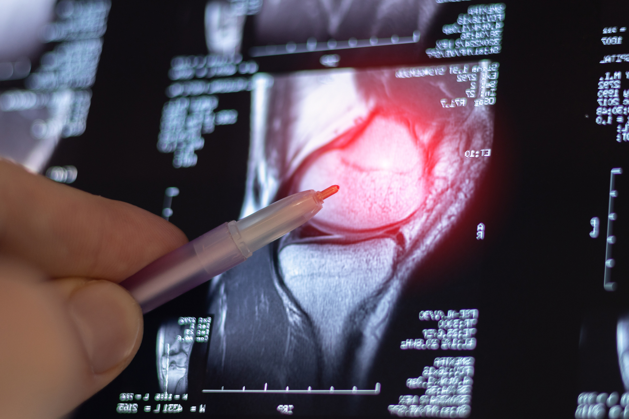
A cartilage defect is a localized area of damage to the smooth, protective surface that lines the ends of your bones inside a joint. It’s not the same as general joint wear, and it’s not arthritis. What makes it particularly serious is that cartilage has almost no ability to repair itself. A defect won’t improve on its own, and the symptoms are easy to mistake for something minor. Getting evaluated early is what keeps a manageable problem from turning into a much bigger one.
At a Glance: Cartilage Defects
- A cartilage defect is localized damage to the joint surface, which is distinct from widespread arthritis
- Cartilage has no blood supply, so it can’t repair itself the way bone, skin, or muscle can
- Symptoms like intermittent swelling and a catching or locking sensation are easy to brush off, but often signal real damage
- Defects are graded 1 through 4 based on depth, with Grade 4 meaning the bone is fully exposed
- Catching a defect early opens the door to more treatment options and better outcomes
Why Cartilage Injuries Don’t Heal on Their Own
Most tissues heal because blood rushes to an injury and delivers the immune cells and growth factors that start the repair process. Cartilage doesn’t work that way. Articular cartilage has no blood supply and no lymphatic network. It gets nutrients only through diffusion from synovial fluid, the lubricating fluid inside your joint. The cartilage cells, called chondrocytes, are locked in place and can’t migrate to fill a defect. Without intervention, a cartilage defect stays or gets worse.
How Is a Cartilage Defect Different from Arthritis?
Osteoarthritis involves diffuse, widespread cartilage breakdown across an entire joint, typically developing over many years. A cartilage defect, also called a focal chondral defect, is more specific: a localized hole or lesion in the cartilage surface. It can affect active adults and athletes of any age across the Midlands, often developing after an ACL tear, a knee dislocation, or a series of cumulative smaller impacts.
Focal chondral defects affect 4.2 to 6.2% of the general population under 40 and show up in up to 36% of athletes. That distinction matters because a focal defect can often be repaired. Widespread arthritis can’t. Getting the right diagnosis is the difference between those two paths.
What Does Full-Thickness Cartilage Defect Mean?
Cartilage defects are graded 1 through 4 based on how deep the damage goes. Grade 1 is surface softening. Grade 2 involves partial-thickness damage that doesn’t yet reach the bone. Grade 3 is deeper damage extending toward the bone. Grade 4 is a full-thickness defect, meaning the cartilage is completely worn through and the bone underneath is exposed.
A full-thickness cartilage defect carries the greatest risk of progressing to widespread joint degeneration without treatment, and it’s the classification most associated with needing more involved intervention down the road.
The Symptoms Worth Paying Attention To
Cartilage defects catch people off guard because the symptoms aren’t always dramatic and don’t always match the severity of the damage. A Grade 4 defect can produce mild, intermittent symptoms for months or years before someone seeks care.
Recurring swelling after activity is often the only symptom in early or moderate defects. A catching or locking sensation when bending the knee, grinding or clicking during movement, pain with stairs or squatting, and an occasional feeling of the knee giving way are all warning signs worth noting. Pain that comes and goes is easy to rationalize away. Most people assume they strained something and move on. The underlying damage doesn’t move on with them.
What Does Cartilage Defect Treatment Look Like?
Treatment depends on defect size, depth, and how long it’s been present. Smaller defects caught early have the most options.
Non-surgical approaches are usually the starting point: physical therapy, anti-inflammatory medications, corticosteroid injections, viscosupplementation (hyaluronic acid), and regenerative options like platelet-rich plasma (PRP) therapy.
For defects that don’t respond, surgical options include:
- Microfracture: your surgeon creates small holes in the bone beneath the defect to stimulate a healing response. This works best for smaller lesions.
- Osteochondral autograft (mosaicplasty): your surgeon moves healthy cartilage and bone plugs from a low-demand area of your own joint to fill the defect.
- Osteochondral allograft: donor cartilage and bone tissue fills the defect, used for larger areas of damage.
- MACI (Matrix-Associated Autologous Chondrocyte Implantation): your surgeon collects your own cartilage cells, grows them in a lab, and places them onto a collagen scaffold. Orthopedic specialists generally prefer this option for larger lesions in younger, active patients.
Research published in PMC found that 52 to 96% of patients report good to excellent outcomes when the right procedure is matched to the right defect.
Why Timing Makes a Bigger Difference Than You’d Think
An untreated cartilage defect doesn’t stay static. Research published in a separate PMC study found that both partial and full-thickness defects significantly increase the risk of further cartilage damage in neighboring areas of the joint. Left untreated, a focal defect can progress to early-onset osteoarthritis and eventually lead to knee replacement surgery.
Earlier treatment means more options and better outcomes. By the time pain becomes severe enough to prompt an orthopedic visit, the defect has often been present much longer than the patient realized. For a closer look at what cartilage restoration can involve, see our blog on cartilage transplantation.
Learn More About Cartilage Defects
Can a cartilage defect heal on its own?
No. Cartilage has no blood supply, which means it lacks the mechanism most tissues rely on to repair themselves. Without blood vessels delivering immune cells and growth factors to the injury site, there’s no natural repair pathway. Conservative management can reduce symptoms and slow progression, but structural damage doesn’t reverse without intervention.
What causes cartilage defects?
Cartilage defects can develop from a single traumatic event like an ACL tear, a knee dislocation, or a direct blow to the joint. They can also build gradually from repetitive impacts, which is common in athletes and people with active jobs or lifestyles. Underlying factors like joint instability and lower extremity malalignment can also increase the risk of developing a defect over time.
When should I see a doctor about knee pain that might be a cartilage defect?
If you’re experiencing recurring swelling after activity, a catching or locking sensation, grinding during movement, or pain with stairs and squatting that doesn’t fully resolve with rest, those symptoms are worth getting evaluated. A dramatic injury isn’t required to justify an appointment. An orthopedic specialist can examine your knee and order the right imaging to determine what’s going on. The earlier a defect is identified, the more treatment options remain available.
The Sooner You Know, the More You Can Do
Cartilage defects are one of those conditions where waiting genuinely costs you. The tissue can’t heal itself, the damage tends to progress, and treatment options available early simply aren’t available once a defect has grown. If something in your knee feels off, it’s worth finding out what it is.
Get Your Knee Evaluated at Palmetto Bone & Joint
If you’re dealing with unexplained knee swelling, a catching sensation, or pain that keeps coming back, the orthopedic and sports medicine team at Palmetto Bone & Joint is here to help. Call us at (803) 902-3169 or contact us online to schedule an evaluation at our Chapin, Irmo, or Newberry locations.
Related Posts

Why Fall Is the Smartest Time to Schedule Your Joint Replacement Surgery
If you’ve been managing joint pain through another summer and telling yourself you’ll deal with…

Laminoplasty vs. Spinal Fusion: How to Know Which Surgery Is Right for You
A hand that won’t quite cooperate, a grip that keeps slipping, numbness that spreads instead…

Carpal Tunnel vs. De Quervain’s: How to Tell Which One Is Causing Your Hand Pain
Carpal tunnel syndrome and De Quervain’s tenosynovitis both cause hand and wrist pain, and they’re…